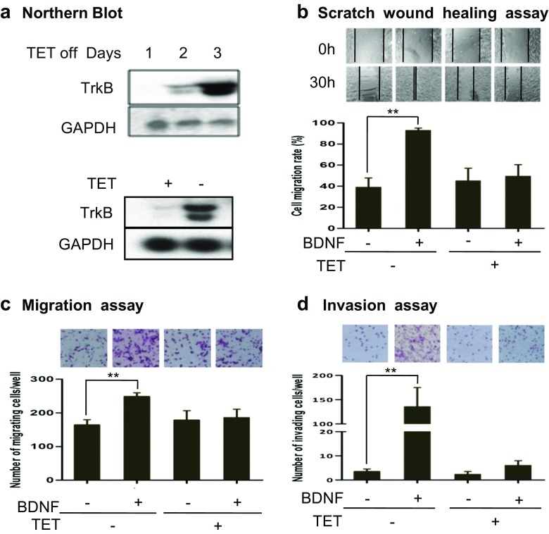
Fig. 1.
The roles of BDNF/TrkB in TB3 cell migration and invasion in vitro. a TET regulated TrkB-expressing cell line. Normally, TB3 cells were cultured with TET (1 μg/ml). To induce the TrkB expression, TET was removed from culture media for 1, 2, and 3 days, and then TB3 cells were harvested and Northern blot was performed. A comparison of TrkB expressions in TB3 cells with or without TET (1 μg/ml, 3 days) was also done. b, c The role of BDNF/TrkB on TB3 cell migration. Scratch wound healing assay was performed. Cell migration was photographed at 10× magnification at 0 h and 30 h after treated with BDNF. The wound healing width was measured by Image-Pro Plus software. The cell migration rate was calculated as described in “Materials and methods” section. Bars, SD. **P < 0.01, BDNF-treated vs. control (b). Migration assay was done as described in “Materials and methods” section. Representative fields of migrating cells under microscope were shown upper of the figure. The cells that migrated to the underside of the inserts were counted, and Student’s t test was done. Bars, SD. **P < 0.01, BDNF-treated vs. control (c). d The role of BDNF/TrkB on cell invasion. Invasion assay was performed as described in “Materials and methods” section. Representative fields of invading cells under microscope were shown upper of the figure. The cells that invaded to the underside of the inserts were counted, and Student’s t test was done. Bars, SD. **P < 0.01 BDNF-treated vs. control. The experiments were repeated three times

