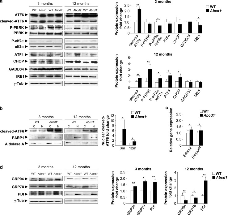
Fig. 2.
UPR induction in the X-ALD mouse model. a Representative immunoblots of ER stress sensors full-length ATF6, cleaved-ATF6, total PERK, P-PERK, eIF2α, P-eIF2α, ATF4, CHOP, GADD34 and IRE1 in the spinal cord tissue of Abcd1 − mice and age-matched wild type (WT) mice at 3 and 12 months of age. The histograms on the right show the cleaved-ATF6, P-PERK, ATF4, CHOP, GADD34 and IRE1 levels normalized relative to γ-Tub and the P-PERK/PERK and the P-eIF2α/eIF2α ratios relative to their respective WT values. b The nuclear localization of cleaved-ATF6 in WT and Abcd1 − mice at 3 and 12 months of age. PARP1 was used as the control for the nuclear fraction (N) and aldolase A was used for the cytoplasmic fraction (C). The histograms on the right show the cleaved-ATF6 levels relative to WT values in the nuclear fractions. c Real-time RT-PCR analyses of Edem2 and Herpud1 mRNA at 12 months in Abcd1 − mouse spinal cords. d Immunoblots of GRP94 and GRP78 chaperones and PDI in the spinal cord tissue of Abcd1 − mice and age-matched wild type (WT) mice 3 and 12 months of age. The histograms on the right show the GRP94 and GRP78 chaperones and PDI levels normalized to WT mice and normalized relative to the γ-Tub. Values are expressed as the mean ± SD (n = 10 samples per genotype and condition in a, c and d; n = 6 samples per genotype and condition in b; *P < 0.05, **P < 0.01 and ***P < 0.001, Student’s t test)

