
Fig. 4.
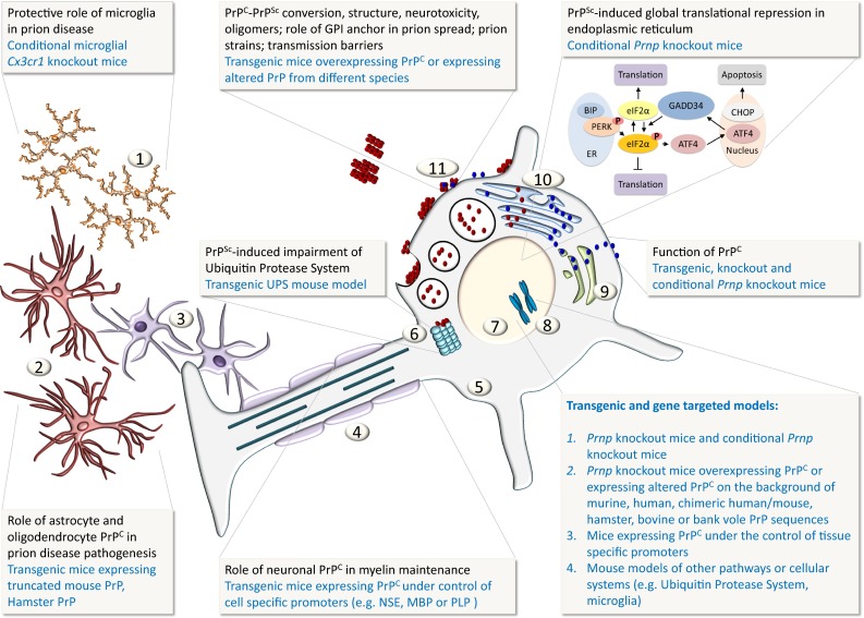
Schematic representation of the cellular and sub-cellular compartments and how PrP function and disease mechanisms are investigated with animal models. Key: 1 microglia, 2 astrocytes, 3 oligodendrocytes, 4 myelinated axon, 5 neuronal cytoplasm, 6 ubiquitin protease system (UPS), 7 neuronal nucleus, 8 chromosomes, 9 Golgi complex, 10 Endoplasmic reticulum (ER), 11 blue globules represent native cellular prion protein PrPC, red globules represent misfolded prion protein PrPSc