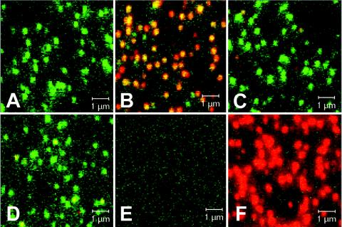
FIG. 4.

Autofluorescence of eGFP-P-containing SAD ΔP virions and coimmunostaining for viral proteins. (A to D) Density gradient-purified SAD ΔP virions complemented with eGFP-P were adhered to glass coverslips and directly analyzed by confocal laser-scanning microscopy (A) or were immunostained (red fluorescence) with sera recognizing RV G (B), M (C), or N (D). (E) Negative control: no virus, anti-RV G immunostain. (F) SAD L16 virions stained with anti-G antibodies. Overlays of the eGFP-P fluorescence (green) and of the immunostainings (red) are shown.
