
FIG. 4.
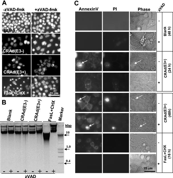
Lack of apoptotic features in H460 cells undergoing cell death triggered by CRAd(E3+) and CRAd(E3−). (A) H460 cells were infected with both CRAds at an MOI of 25 alone or in combination with 100 μM zVAD-fmk for 2 days. As a control, cells were exposed for 16 h to FasL+CHX with or without zVAD-fmk. Nuclei were stained with Hoechst 33342 to visualize chromatin condensation, nuclear shrinkage, or fragmentation that are markers for apoptosis by immunofluorescence microscopy. CRAd-infected cells did not display nuclear apoptotic features, in contrast to Fas+CHX-exposed cells that show condensed and fragmented nuclei reversible by zVAD-fmk. (B) DNA fragmentation was also analyzed, revealing no apoptotic DNA laddering in CRAd-infected cells, whereas FasL+CHX treatment results in clear DNA smearing. (C) Annexin V staining caused by PS externalization was examined 1 and 2 days postinfection and in FasL+CHX-treated H460 cells with and without zVAD-fmk. Cells were cotreated with PI to allow the distinction between Annexin V staining in the presence or absence of an intact membrane detected by immunofluorescence microscopy; the phase-contrast image is also shown (Phase). A minor portion of CRAd-infected cells were PI positive (indicated by arrows). Annexin V staining is caspase independent in CRAd-infected cells and caspase dependent when induced by FasL+CHX. Results are shown for CRAd(E3+)-infected cells; similar findings were obtained for CRAd(E3−)-infected cells.