
FIG. 1.
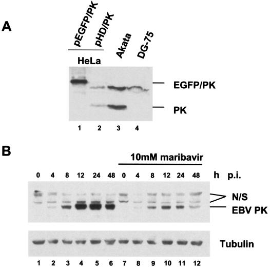
EBV PK expression during viral reactivation in Akata cells. (A) Equal amounts of whole-cell lysates from the HeLa cells transfected with pEGFP/PK or pHD/PK, Akata cells (24 h after viral reactivation), and DG-75 cells were subjected to SDS-PAGE, followed by immunoblotting with the affinity-purified anti-PK antibody. (B) Akata cells were collected at different time points after viral lytic reactivation in the presence or absence of maribavir, and equal amounts of the whole-cell lysates were analyzed by immunoblotting them with anti-PK antibody. Lanes 1 to 6, no maribavir; lanes 7 to 12, samples treated with 10 μM maribavir. Lanes 1 and 7 are latent Akata cells. Nonspecific bands detected by the antibody are marked N/S.