
FIG. 3.
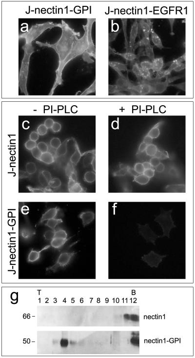
Expression of nectin1-GPI and nectin-EGFR1. Nectin1-GPI is anchored to the plasma membrane through the GPI lipid moiety and is sorted to lipid rafts. Images show J-nectin1-GPI cells (a, e, and f), J-nectin1-EGFR1 cells (b), and J-nectin1 cells (c and d). (a and b) Cells were fixed with methanol and reacted with MAb R1.302 to nectin1, followed by secondary FITC-conjugated antibody. Fluorescence localized at the level of the plasma membrane and intracellularly. (c to f) Cleavage of nectin1 from nectin1-GPI with PI-PLC. J-nectin1 cells (c and d) and J-nectin1-GPI cells (e and f) were mock treated (c and e) or treated with PI-PLC (d and f). Cells then were fixed with paraformaldehyde and reacted with MAb R1.302, followed by secondary FITC-conjugated antibody. (g) Flotation of nectin1-GPI in raft-containing low-density sucrose fractions. J-nectin1-GPI and J-nectin1 cells were solubilized in Triton X-100 and fractionated on sucrose gradients. The proteins were separated by SDS-PAGE, and nectin1 was visualized by Western blotting with MAb CK6 directed to the nectin1 ectodomain, followed by peroxidase-conjugated anti-mouse antibody and ECL. Numbers above panels indicate fractions collected from top (T) to bottom (B); numbers at left indicate the apparent molecular weight.