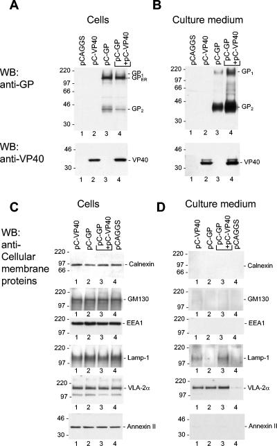
FIG. 1.

Detection of GP, VP40, and cellular membrane proteins in cell lysate and cellular supernatant by immunoblot analysis. (A to D) 293 cells were transfected with the plasmids encoding the proteins indicated above the lanes. At 48 h posttransfection, the cells and particulate material of cellular supernatant were harvested, diluted as described in Materials and Methods, and subjected to SDS-PAGE (12% polyacrylamide). The positions of the immature form of GP located in the ER (GPER), GP1, GP2, VP40, and cellular membrane proteins are indicated. (A and C) Cell lysate; (B and D) supernatant.
