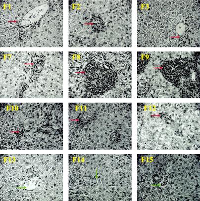
FIG. 3.

Histopathology of ferret livers following immunization and SARS-CoV challenge. Representative pictures were taken at 27 to 29 days postinfection (ferrets [F] 1 to 3, 7 to 9, and 10 to 12) or no infection (naive ferrets 13, 14, and 15; exposed to neither MVA nor SARS-CoV) at ×40 magnification. Perivascular mononuclear infiltrates (red arrows) were present in all livers from ferrets infected with SARS-CoV, ranging from mild (ferrets 1, 2, 3, 10, 11, and 12) to severe (ferrets 7, 8, and 9) lesions. In addition, intralobular infiltration of mononuclear cells was extensive in livers from ferrets 7, 8, and 9. No significant liver lesions were found in naive ferrets (ferrets 13, 14, and 15, which did not receive MVA or SARS-CoV and which were used for preliminary examination of ferrets. Green arrows, vein of portal triads.
