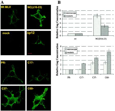
FIG. 3.

Localization and quantitation of mutant Gag proteins by immunofluorescence and immunoblotting. Cells were first transfected with the indicated viral clones and then fixed and stained for detection of CA as described in Materials and Methods. (A) Immunofluorescence images. Bar, 10 μm. (B) Relative concentrations of anti-CA-reactive material in the cells, measured either by integrating immunofluorescence intensity over the cells as described in Materials and Methods (microscope) or by immunoblotting (Western). wt, wild type.
