
FIG. 1.
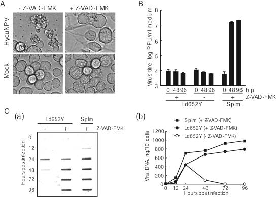
Cytopathology, BV yields, and viral DNA accumulation in HycuNPV-infected and Z-VAD-FMK-treated Ld652Y cells. Monolayer cultures of Ld652Y cells were infected with HycuNPV at an input MOI of 5 PFU/cell and were cultured in medium only or in medium containing 20 μM Z-VAD-FMK. (A) Cytopathology of HycuNPV-infected and Z-VAD-FMK-treated Ld652Y cells at 96 hpi. Mock-infected and Z-VAD-FMK-treated Ld652Y cells were incorporated as controls. (B) BV yields from HycuNPV-infected and Z-VAD-FMK-treated Ld652Y cells were determined by plaque assay on SpIm cells. Vertical bars indicate standard deviations of averages from three determinations. (C) Slot blot hybridization analysis of viral DNA in HycuNPV-infected and Z-VAD-FMK-treated Ld652Y cells. The viral DNAs were blotted onto a Hybond-N+ membrane and were hybridized with fluorescein-labeled hycu-iap3 gene probe. The probe was visualized by gene images (a) and was quantified with a Lumi Imager by comparing the signal intensities in infected cells with those of serially diluted HycuNPV DNAs of known amount (b). (B and C) HycuNPV-infected and Z-VAD-FMK-treated SpIm cells (conventional host cells) are also shown for comparison.