
FIG. 2.
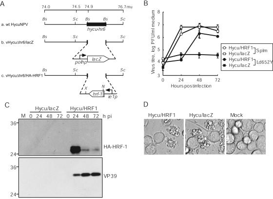
Viral replication in vHycuΔhr6/HA-HRF1- and vHycuΔhr6/lacZ-infected Ld652Y cells. (A) Schematic representation of wt HycuNPV, vHycuΔhr6/lacZ, and vHycuΔhr6/HA-HRF1. The schematics represent the 74.0- to 76.7-mu region of wt HycuNPV (a) as well as the genetic modification of vHycuΔhr6/lacZ (b) and vHycuΔhr6/HA-HRF1 (c), in which hycu-hr6 was replaced by the lacZ gene under the control of the SeMNPV polyhedrin promoter (polhp) and the HA tag-fused hrf-1 gene under the control of the hycu-ie1 promoter (ie1p), respectively. Bs, BstXI; N, NcoI; Sc, SacII; X, XbaI. (B) BV yields in culture medium determined by plaque assay on SpIm cells. (C) Accumulation of HA-HRF-1 and VP39 in infected cells monitored by immunoblotting using antisera against HA and VP39, respectively. (D) Photographs of infected cells at 72 hpi. (B, C, and D) Monolayer cultures of Ld652Y cells were infected with vHycuΔhr6/HA-HRF1 (Hycu/HRF1) and vHycuΔhr6/lacZ (Hycu/lacZ) at an input MOI of 5 PFU/cell. Vertical bars in panel B represent standard deviations of averages from three determinations. The numbers on the left of panel C indicate molecular masses (kilodaltons) of marker proteins (BenchMark Prestained Protein Ladder; Invitrogen). M, mock-infected cells.