
FIG. 3.
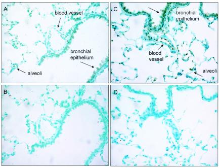
Immunohistochemical analysis of EGFP expression in 0.5-μm sections of rat lung, 42 days after AAV[2/2]CMV-EGFP or AAV[2/5]CMV-EGFP vector-mediated endotracheal administration. Positive cells are present in alveolar and airway epithelia as well as om vascular endothelia when AAV[2/2] and AAV[2/5] were given, although they are significantly more intense in the AAV[2/5]-treated lung. (A and B) AAV[2/2]CMV-EGFP vector-treated lung sections reacted with rabbit anti-EGFP IgG or the isotype control antibody, respectively. (C and D) AAV[2/5]CMV-EGFP vector-treated lung sections reacted with rabbit anti-EGFP IgG or the isotype control antibody, respectively.