
FIG. 3.
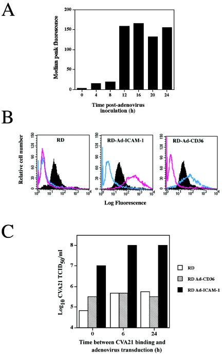
CVA21 induced lytic infection of RD cells following delayed induction of ICAM-1 expression. (A) Time course of ICAM-1 expression following adenovirus transduction. RD cells were induced to express human ICAM-1 by transduction with 2.5 × 10⁶ TCID50 of a recombinant adenovirus containing human ICAM-1 cDNA. Cells were assessed by flow cytometry for ICAM-1 expression at various times after adenovirus inoculation with the anti-ICAM-1 domain monoclonal antibody (WEHI). (B) Flow-cytometric analysis of RD cells showing surface expression of DAF, ICAM-1, and CD36 24 h following mock transduction or transduction with recombinant adenoviruses containing human ICAM-1 or CD36 cDNA. The solid histograms represent DAF expression, while the pink histograms represent ICAM-1 expression and the blue histograms represent CD36 expression. The recombinant adenoviruses containing ICAM-1 cDNA or CD36 cDNA were constructed with an Adeno-quest kit (Quantum Biotechnologies Inc.) in accordance with the manufacturer's instructions. (C) CVA21 lytic infection of RD cells via the delayed expression of ICAM-1. Monolayers of DAF-expressing RD cells in six-well culture plates were infected with the prototype strain of CVA21 (multiplicity of infection = 1.0 TCID50) for 1 h at 37°C. Following removal of non-DAF-bound CVA21 virions by four washes with serum-free Dulbecco's modified Eagle medium (DMEM), cell monolayers were transduced with 2.5 × 10⁶ TCID50 of a recombinant adenovirus containing ICAM-1 or CD36 cDNA immediately or at 6 and 24 h after CVA21 inoculation. At 6 and 24 h after CVA21 inoculation, cell monolayers were washed with serum-free DMEM prior to adenovirus transduction. Following adenovirus transduction, cell monolayers were incubated at 37°C for 24 h, at which time cell supernatants were harvested. RD cells that were inoculated with CVA21 but mock transduced served as background cell controls. Levels of progeny CVA21 in the cell supernatants were determined by lytic-end point dilution on 96-well monolayers of ICAM-1-expressing RD cells. Cell survival from quadruplicate wells was quantitated by staining with a crystal violet-methanol solution, and the relative absorbance of stained cell monolayers was read on a multiscan enzyme-linked immunosorbent assay plate reader (Flow Laboratories) at 540 nm. Fifty percent end point titers were calculated by the method of Reed and Muench (21), where a well was scored as positive if the absorbance was less than the no-virus control minus three standard deviations.