
FIG. 4.
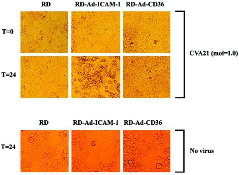
CVA21-induced lytic infection of RD cells transduced with ICAM-1- or CD36-expressing adenovirus. DAF-expressing RD cells were infected with the prototype strain of CVA21 (multiplicity of infection [moi] = 1.0 TCID50) for 1 h at 37°C. Following removal of non-DAF-bound CVA21 virions by four washes with serum-free Dulbecco's modified Eagle medium, cell monolayers were transduced with 2.5 × 10⁶ TCID50 of a recombinant adenovirus (Ad) containing ICAM-1 or CD36 cDNA. Following incubation for 24 h at 37°C, cell monolayers were microscopically examined for cell lysis. Photomicrographs were taken at a magnification of ×200. Time (T) zero represents cell monolayers immediately after adenovirus transduction, while time 24 represents cell monolayers 24 h following adenovirus transduction.