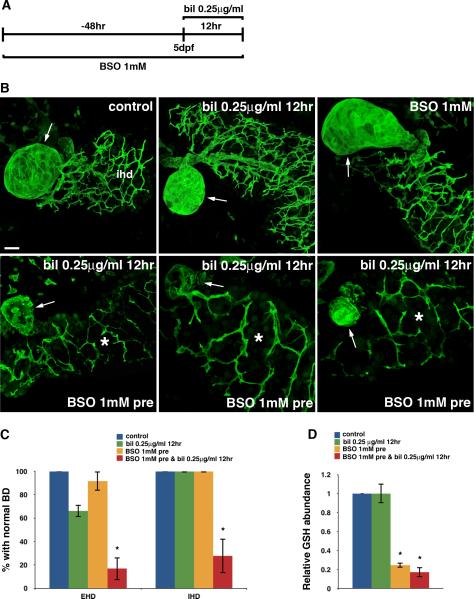
Fig. 3. GSH depletion sensitizes cholangiocytes to biliatresone.
(A) Experimental scheme depicting treatment of 3 dpf wild-type larvae with BSO (1 mM) for 48 hr prior to low-dose biliatresone exposure at 5 dpf (0.25 μg/ml, 12 hr). (B) Confocal projections through the livers of 5 dpf larvae immunostained with anti-Annexin A4 antibody. The top three images show control, biliatresone-treated, and BSO-treated larvae. Morphology of the gallbladder (arrow) and IHD is normal in the control larva and larvae treated with low-dose biliatresone or BSO. In contrast, significant destruction of both the EHD (gallbladder-arrow) and IHD (*) is noted in biliatresone-treated larvae preconditioned with BSO (bottom panels). Scale bar, 20 μm. (C) Percentage of larvae exposed to biliatresone or biliatresone and BSO with normal EHD and IHD morphology. *p < 0.001 compared to control larvae. (D) Hepatic GSH levels in low-dose biliatresone-treated larvae with and without BSO pretreatment. *p < 0.001 compared to control larvae. All data represent mean of at least three experiments with 5-10 larvae per condition for each experiment. Error bars, SEM. Abbreviations: bil, biliatresone; BD, bile duct; BSO, buthionine sulfoximine; EHD, extrahepatic bile duct; IHD, intrahepatic bile ducts.

