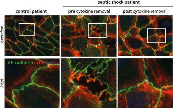
Fig. 1.

Endothelial phenotype with respect to barrier function. Fluorescence immunocytochemistry staining for vascular endothelial (VE)-cadherin (green), F-actin (red), was performed on confluent human umbilical vein endothelial cells (HUVECs) as described before [5]. Cells were treated for 30 min with media supplemented with 5% serum from an individual with septic shock before (2nd row) and after cytokine removal (3rd row); 5% healthy human serum served as a control (1st row). Scale bar 10 μm
