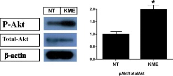
Fig. 1.

Activation of the Akt signaling pathway by KME (Left) Relative phosphorylation of Akt (P-Akt) is shown. The concentration of KME was 100 μg/ml. (Right) The relative amount of phospho-Akt is shown. All the samples were blotted against β-actin as a loading control. NT: non-treated cells. P-values of < 0.05 and < 0.01 are indicated by * and **, respectively
