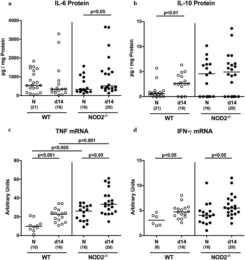
Fig. 5.

Colonic cytokines in C. jejuni infected conventionally colonized NOD2−/− mice. Wildtype (WT; white circles) and NOD2−/− mice (black circles) were perorally infected with C. jejuni strain 81–176 on three consecutive days (d0, 1 and 2). a IL-6 and b IL-10 protein concentrations were measured in colonic ex vivo biopsies at day 14 post infection. In additon, large intestinal c TNF and d IFN-γ mRNA expression levels were determined by real time PCR and expressed as arbitrary units (fold expression). Naive (N) mice served as uninfected controls. Medians (black bars), levels of significance (p value) determined by Mann–Whitney U test and numbers of analyzed animals (in parentheses) are indicated. Data were pooled from four independent experiments
