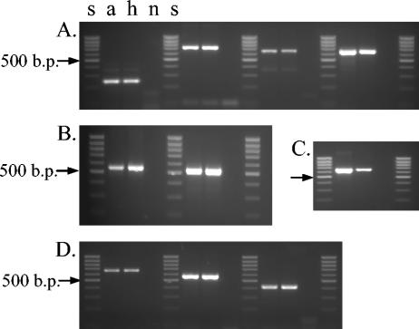
FIG. 5.
MIRU-VNTR analysis of batches a and h of M. tuberculosis strain 98/426 with the primers described by Supply et al. (28). Amplification products were checked on a 2.5% agarose gel, run for 90 min at 45 V, and examined under UV light after staining with ethidium bromide. Panels: A, loci 4, 16, 10, and 31 (left to right); B, loci 26 and 40; C, locus 39; D, loci 2, 23, and 27. Lanes s, 100-bp ladder (Biotools, B&M Labs) used as a DNA molecular size marker; lane n, negative control.

