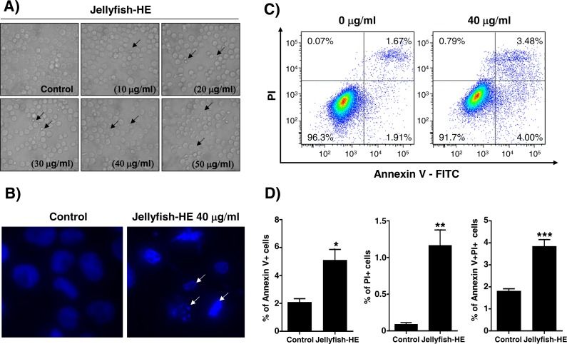
Figure 3. Jellyfish hexane extract induces apoptosis in K562 cells.
(A) Morphology. K562 cells were treated for 24 h with various concentrations of Jellyfish-HE. Phase contrast microscopic observation was been made using a Nikon TMS (Tokyo, Japan). Arrows indicate apoptotic bodies, which are characteristic of cell death. After 24 h incubation with or without Jellyfish-HE, nuclear fragmentation was stained by DAPI for 10 min at 37 °C by fluorescence microscopy (Zeiss Axioskop 2 microscope) (B). Arrows indicate fragmented nuclei. To observe apoptotic cell death in the earlier stages of treatment with Jellyfish-HE, cells were treated with Jellyfish-HE for 8 h. Then, apoptotic cells were detected by PI and Annexin V double staining by flow cytometry (FACS Canto II) (C) and quantitative analysis (D). * P < 0.05, ** P < 0.005 and *** P < 0.0005 vs. control (untreated).

