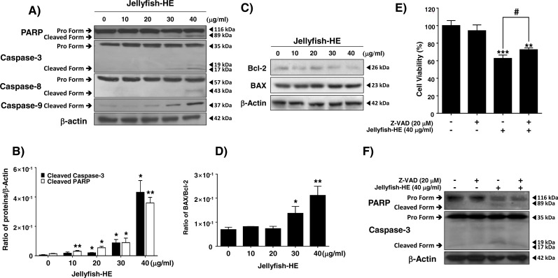
Figure 4. Jellyfish hexane extract induces apoptosis via extrinsic and intrinsic pathways in K562 cells.
(A) After treatment of K562 cells with various concentrations (0, 10, 20, 30, and 40 mg/ml) of Jellyfish-HE for 24 h, the expression levels of PARP, caspase-3, caspase-8 and caspase-9 were analyzed by immunoblotting with antibodies specific for PARP, caspase-3, caspase-8, and caspase-9. (B) The ratio of each protein to β-actin was calculated using Image J software. (C) After treatment with Jellyfish-HE at various concentrations for 24 h, levels of Bcl-2 and BAX proteins were analyzed by immunoblotting and the BAX/Bcl-2 ratio was quantified by densitometry (D). K562 cells were treated with 40 mg/ml Jellyfish-HE for 12 h in the presence or absence of Z-VAD and then (E) cell viability was measured by an MTT assay and analyzed by immunoblotting (F) with antibodies specific for PARP and capase-3. b-Actin was used as a loading control. * P < 0.05, ** P < 0.005 and *** P < 0.0005 vs. control (untreated). # P < 0.05 vs. treatment with Jellyfish-HE.

