
FIG. 4.
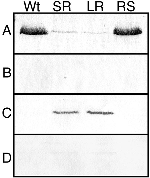
Anti-CwhA immunoblot of cellular fractions from KM'92 isolates. Proteins from exponential-phase (OD600 = 0.7) wild-type (Wt), SR, LR, and RS cells were fractionated as described in Materials and Methods into (A) extracellular supernatant, (B) cell wall, (C) cell membrane, and (D) cytoplasm. Equivalent loadings (50 μg of protein) from the extracellular supernatant, cell membrane, and cytoplasm, and 20 μl, corresponding to one-fifth of the total cell wall fraction, were separated by SDS-PAGE on 10% acrylamide gels. The separated proteins were electrotransferred to nitrocellulose and probed with the PepD-CwhA monoclonal antibody as described in Materials and Methods. The antigenic band detected in the preparations had an apparent molecular size of ≈55 kDa. Stationary-phase cells gave identical results.