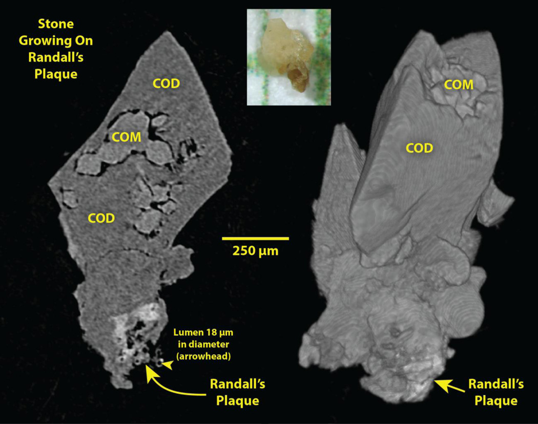
Figure 1.
Stone growing on Randall’s plaque, plucked from the papilla of a patient during ureteroscopic removal of renal stones. Photo, top, on mm paper. Left: Micro CT slice of stone. Right: Surface rendering of micro CT image stack. COM: calcium oxalate monohydrate. COD: calcium oxalate dihydrate.

