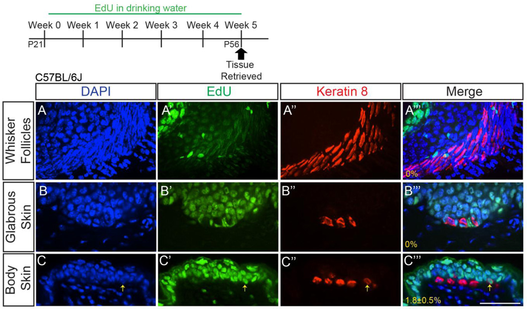
Figure 2. Few adult Merkel cells are formed, and only within touch domes.
Sectioned whisker follicles (z-stack projection; A–A’’’), glabrous forepaw skin (single z-slice; B–B’’’), and back skin (single z-slice; C–C’’’) from female P56 C57BL/6J mice that received 0.2mg/mL EdU in their drinking water for five weeks. Tissues were processed for EdU (A’, B’, C’; green) and K8 immunostaining (A’’, B’’, C’’; red). Yellow arrow (C–C’’’) indicates a K8+EdU+ cell. Percentages of K8+ cells that were EdU+ are shown (A’’’–C’’’) (n=3 mice). Scale bar: 50µm.

