Figure 1.
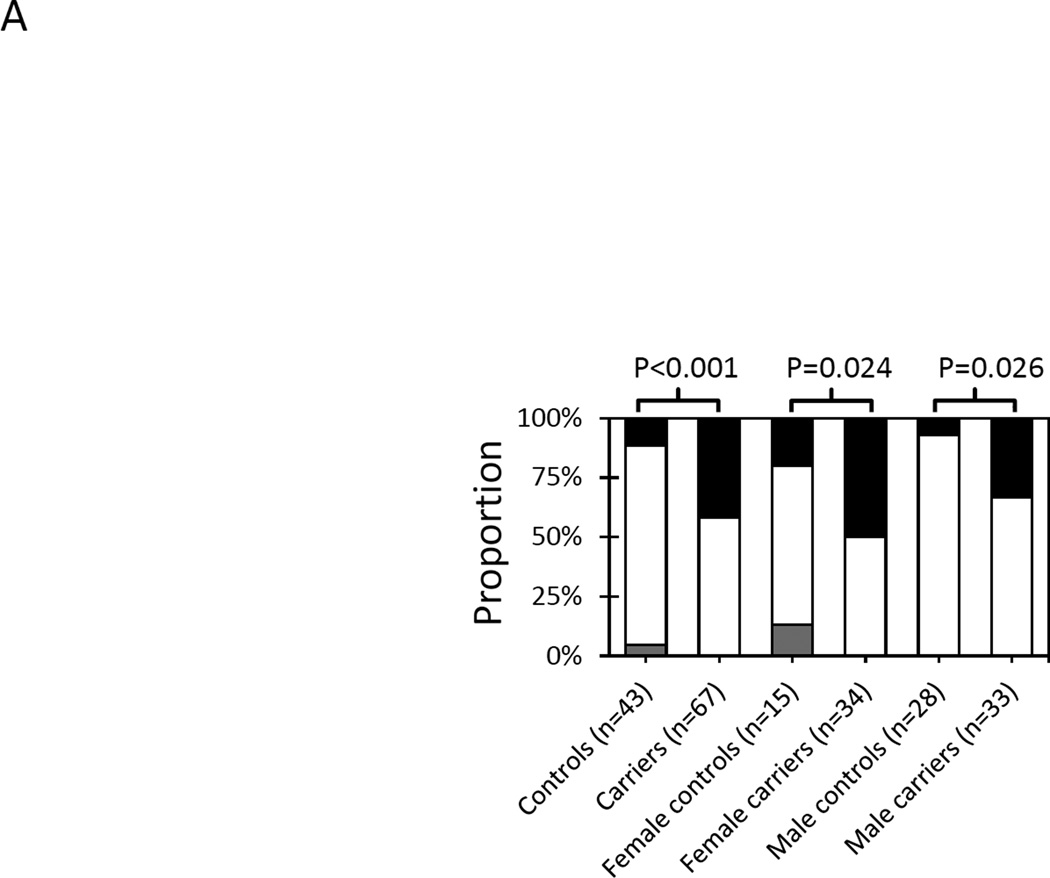
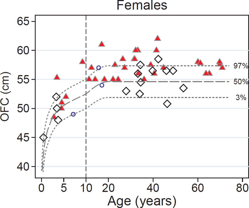
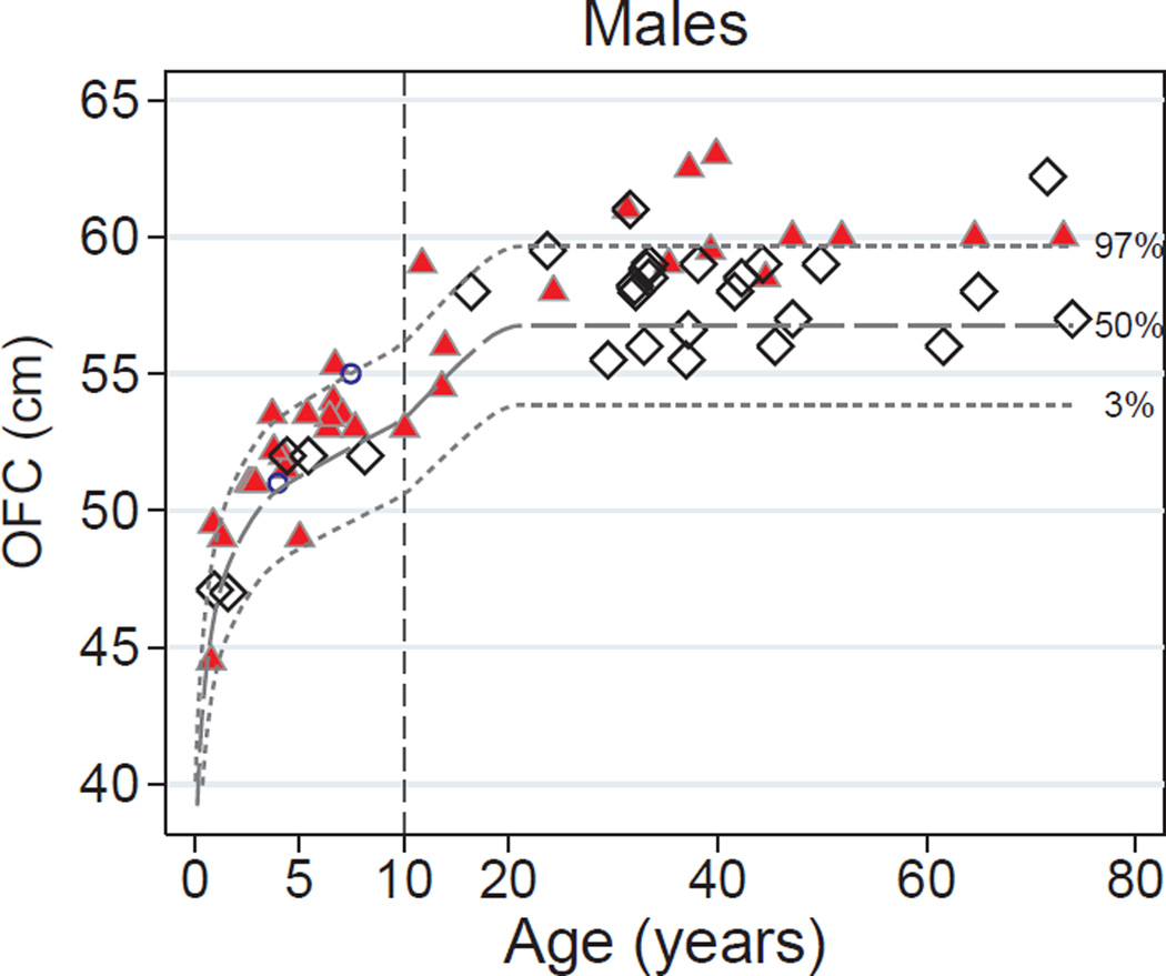
Occipital-frontal circumference-for-age in the DICER1 syndrome. (A) Abnormal OFC-for-age. Proportions between the 3rd and 97th centiles (white), below the 3rd centile (gray), or above the 97th centile (black) in DICER1 mutation carriers and family controls. P values are for Fisher’s exact test of differences between groups. (B) Females. Red triangles indicate DICER1-mutation carriers. White diamonds represent family controls. Blue circles represent girls without a detectable germline DICER1 mutation but who harbor a DICER1-associated tumor (7-year-old: type II PPB; 15.5-year-old: Sertoli-Leydig cell tumor; 17-year-old: type II PPB). The dashed lines indicate the 97th, 50th, and 3rd centiles of OFC-for-age reported in Rollins, 2010. The vertical dashed line at age 10 years indicates a change in the scale of the x-axis to allow for better resolution of children’s values. (C) Males. Blue circles represent boys without a detectable germline DICER1 mutation but who harbor at least one DICER1-associated tumor (4-year-old: type I PPB and cystic nephroma; 7.7-year-old: type II PPB only). OFC = occipital-frontal circumference.


