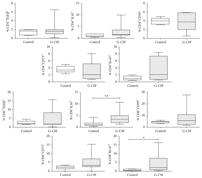
Figure 3.
Cell viability and anti-inflammatory molecules in healthy donors mobilized with G-CSF. Determination of TGFβ, IL-10, CD39, CD73, and Ki-67 in peripheral blood CD4+and CD8+ cells from a control group (n = 6) and a group of G-CSF-mobilized donors (n = 8). Box plots show population distribution and whiskers denote one standard deviation. A significant increase is observed in the number of CD8+IL-10+ and CD8+Ki67+ cells; the change in CD8+CD73+ cells is not significant, but a marked tendency to increase is shown (p = 0.06); no changes or tendencies are seen in marker expression in CD4+ cells. ∗p < 0.05; ∗∗p < 0.01.

