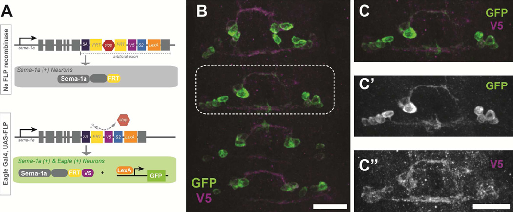
Figure 3. Sema-1a protein is expressed on Eg axons.
An artificial exon inserted into the endogenous locus for sema-1a allows for tissue specific labeling of endogenous Sema-1a expression. (A) Schematic of sparse labeling strategy adapted from Pecot, et al. 2013. In the presence of FLP recombinase, Sema-1a becomes tagged with a V5 epitope and LexA driven membrane bound GFP labels Sema-1a positive cells. (B–C) An early stage 15 embryo carrying the artificial exon, egGal4, UAS-FLP and LexAop-myrGFP stained with anti-GFP (green) and anti-V5 (magenta) antibodies. (B) Eagle neurons endogenously express Sema-1a during midline crossing. Scale bar represents 15µm (C) Magnification of the boxed region in B. (C’) GFP only staining shows two EW axons crossing the midline (C”) V5 staining reveals that Sema-1a protein is expressed throughout the growing axon. Scale bar represents 15µm

