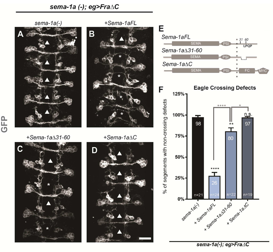
Figure 4. Sema-1a expression rescues midline crossing cell autonomously.
(A–D) Stage 15–16 embryos of the indicated genotypes carrying eg-GAL4, UAS-FraΔC and UAS-CD8 GFP transgenes, stained with anti-GFP (green) antibodies to reveal cell bodies and axons of the eagle neurons (EG and EW). Arrowheads indicate segments with non-crossing EW axons and asterisks indicate rescued EW crosses. Scale bar represents 15µm (D). (A) In sema-1a null embryos expressing FraΔC, EW axons fail to cross the midline in 98% of segments (arrowheads). (B) Expression of a full-length Sema-1a transgene strongly rescues these defects (asterisk), with only 26% non-crossing (arrowheads). (C) In contrast, a Sema-1a transgene lacking a small region of the cytoplasmic domain (from aa31–60) rescued the defects to a much lesser extent (80%). (D) Expression of a Sema-1a transgene lacking its entire cytoplasmic domain does not significantly rescue crossing defects. (E) Diagram of transgenic rescue constructs with relevant cytoplasmic regions labeled. (F) Quantification of EW midline crossing defects in the genotypes shown in (A–D). Data are represented as mean+SEM. n, number of embryos scored for each genotype. Significance was assessed by multiple comparisons using ANOVA (****p<0.0001).

