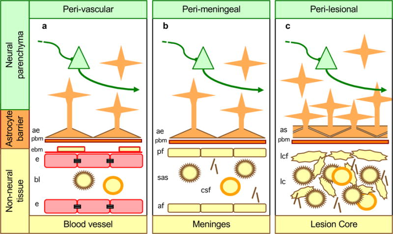
Figure 1. Astrocytes form borders (glia limitans) that serve as functional barriers at interfaces between non-neural tissue and CNS neural parenchyma along blood vessels (a), meninges (b) and tissue lesions (c).

a| Along blood vessels, astrocyte endfeet and parenchymal basement membrane (PBM) present diverse molecular cues that constitute part of the multiple functional barriers, including endothelia with tight junctions and endothelial basement membrane (EBM), across which leukocytes must be actively recruited to pass from the bloodstream into CNS neural parenchyma21, 23, 24. b| Abutting the meninges, astrocyte endfeet and parenchymal basement membranes present multiple molecular cues that restrict leukocytes in the subarachnoid space (SAS) from passing freely into CNS parenchyma. c| Surrounding tissue lesion cores (which are comprised of non-neural cells including leukocytes), astrocyte scars and parenchymal basement membrane are similar in appearance, organization and function to astrocyte borders lining non-neural cells along meninges and blood vessels, and similarly restrict the entry of leukocytes into adjacent CNS parenchyma.

|
neuron |
|
macrophage |
|
|
T cell | ||

|
astrocyte |
|
collagen |
|
|
astrocyte |
|
tight junction |
