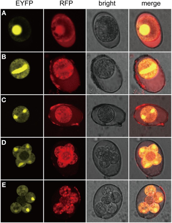
Figure 3.

Expression of exogenous proteins of EmagER during sporogony. Freshly collected oocysts of EmagER were incubated in 2.5% potassium dichromate at 27°C and observed at different time intervals under a confocal microscope (Leica, SP5, Germany). (A) A freshly collected unsporulated oocyst; (B) an oocyst during the process of spindle stage; (C) first nuclear division finished; (D) four separated sporoblasts projected from the central cytoplasmic mass during the second nuclear division; and (E) formation of four spheres of sporocysts and the oocystic residua.
