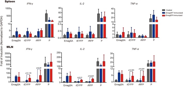
Figure 5.
Transcriptional levels of Th1 cytokines of splenocytes and mesenteric lymph nodes (MLN) of rabbits immunized with EmagWT and EmagER after in vitro stimulation. Three groups of rabbits were inoculated with either 200 oocysts of EmagWT, EmagER, or 200 µl PBS, respectively. Splenocytes and MLN lymphocytes were isolated and stimulated with 20 µg/ml of either EmagWT-soluble antigen, recombinant enhanced yellow fluorescent protein, recombinant red fluorescent protein, or positive stimulus (P, containing 40.5 µM PMA and 670 µM ionomycin). Quantitative real-time PCR was performed using GAPDH as an internal control. Relative gene expression was calculated by the 2−ΔΔCq (Livak) method. All data were presented as mean ± SEM values. Statistical analysis was performed by one-way ANOVA, LSD.

