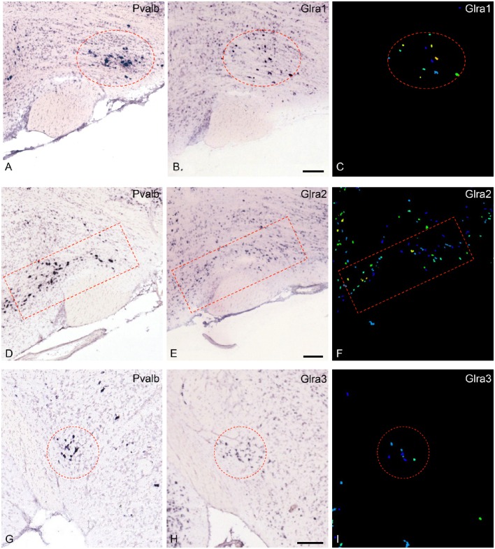
Figure 1.
Expression of the mRNA for the glycine-receptor subunits α-1, α-2 and α-3 in the parvafox nucleus as found in the ABA-ISH. (A,D,G) In situ hybridization-images for Pvalb, downloaded from the ABA website, defining the location of the parvafox nucleus (surrounded by a red circle or rectangle). (B,E,H): The mRNAs for the glycine-receptor subunits α-1, α-2, and α-3 are expressed within the confines of the parvafox nucleus. In (C,F,I) the expression level of Glra1, Glra2, and Glra3 is visualized with pseudocolors. (A–F) are sagittal sections and (G–I) coronal sections. (Image credit: Allen Institute.) Scale bars represent 100 μm.

