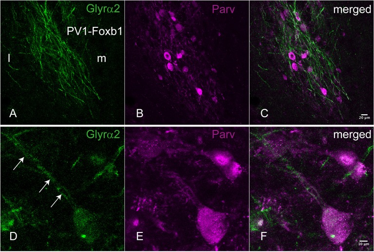
Figure 2.
Immunohistochemical localization of the Glyrα2 receptor in the parvafox nucleus. In overview pictures, the Glyrα2 immunoreactive sites in the hypothalamus are concentrated in the ventrolateral hypothalamus and the mammillary nuclei. At low magnification, a meshwork of Glyrα2 immunoreactive fibers (A) is intermingled with Parv-immunoreactive neurons (B,C). At higher magnification, Glyrα2 immunoreactivity decorates the dendrites (arrows, D) and cell body of Parv-positive neurons (E,F).

