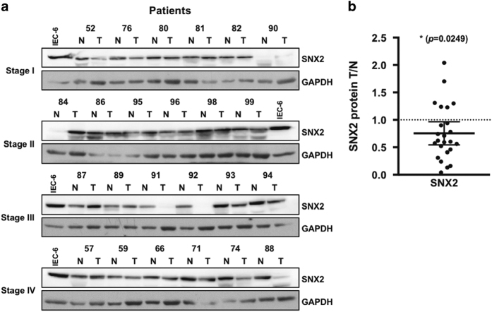
Figure 8.
SNX2 protein levels are decreased in CRC tumors. (a) Samples from our CRC tissue bank were analyzed by immunoblotting for SNX2 protein in adjacent normal (N) and tumor (T) tissues. Glyceraldehyde 3-phosphate dehydrogenase (GAPDH) was used as a loading control to normalize data; the same IEC-6 (intestinal epithelial cell line) cell extract sample was used in all immunoblots to normalize results across different experiments. (b) Densitometric analysis of all CRC stages from (a). The P-value is indicated (t-test). *P<0.05.

