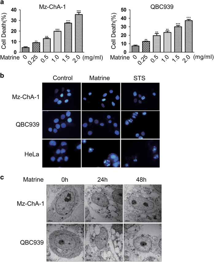
Figure 1.
Matrine-induced non-apoptotic cell death in CCA cells. (a) Matrine-induced cell death in a dose-dependent manner in Mz-ChA-1 and QBC939 cells. Cells were treated with different concentrations (0, 0.25, 0.5, 1.0, 1.5 and 2.0 mg/ml) of matrine for 48 h, then the percentage of cell death was determined with PI staining plus flow cytometry. All data were presented as the mean±S.D. of three independent experiments. Significant differences compared with vehicle controls were indicated as *P<0.05, **P<0.01 and ***P<0.001 (assessed by Student’s t-test). (b) The nuclei of Mz-ChA-1 and QBC939 cells did not display typical apoptotic features under matrine treatment. Cells were treated with matrine (1.5 mg/ml) or vehicle (control) for 48 h, or STS (2 μM) for 12 h, and then subjected to DAPI staining and fluorescent microscope. (c) Accurate morphological analysis of CCA cells treated with matrine using transmission electron microscope. Mz-ChA-1 and QBC939 cells were treated with matrine (1.5 mg/ml) for 24 and 48 h, and then observed and photographed under transmission electron microscope.

