Figure 3.
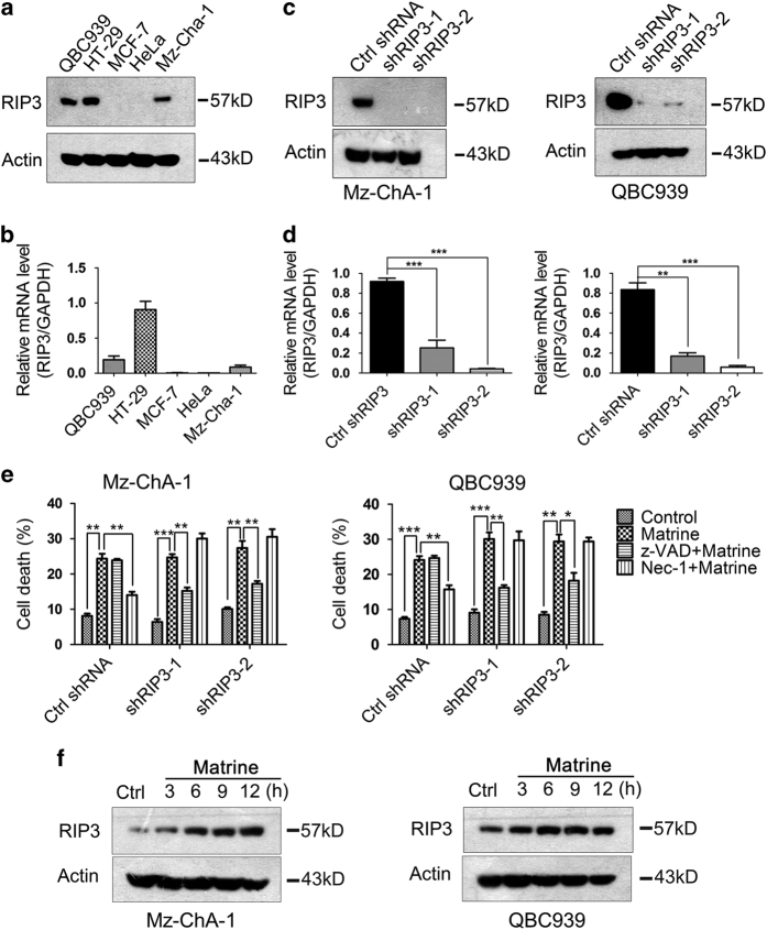
RIP3 was required for matrine to induce necroptosis in CCA cells. (a and b) Endogenous RIP3 expression levels in several tumor cell lines were detected by western blot (a) and real-time PCR (b). (c and d) RIP3 knockdown efficiency in Mz-ChA-1 (Left) and QBC939 (Right) cells was determined by western blot (c) and real-time PCR (d). *P<0.05, **P<0.01 and ***P<0.001 versus control (assessed by Student’s t-test). (e) Mz-ChA-1 and QBC939 cells expressing control or RIP3 shRNA were pre-treated with Nec-1 (20 μM) or z-VAD-fmk (20 μM) for 2 h, and then treated with matrine (1.5 mg/ml) or vehicle for 48 h. After that, the percentage of cell death was determined by PI staining and flow cytometry. Results were presented as the mean±S.D. from three independent experiments. Significant differences were indicated as *P<0.05, **P<0.01 and ***P<0.001 (assessed by Student’s t-test). (f) Matrine increased RIP3 expression levels in Mz-ChA-1 and QBC939 cells. Cells were treated with matrine (1.5 mg/ml) for 0, 3, 6, 9 and 12 h, then lysed and subjected to western blot analysis with anti-RIP3 antibody. β-actin was used as an internal control.

