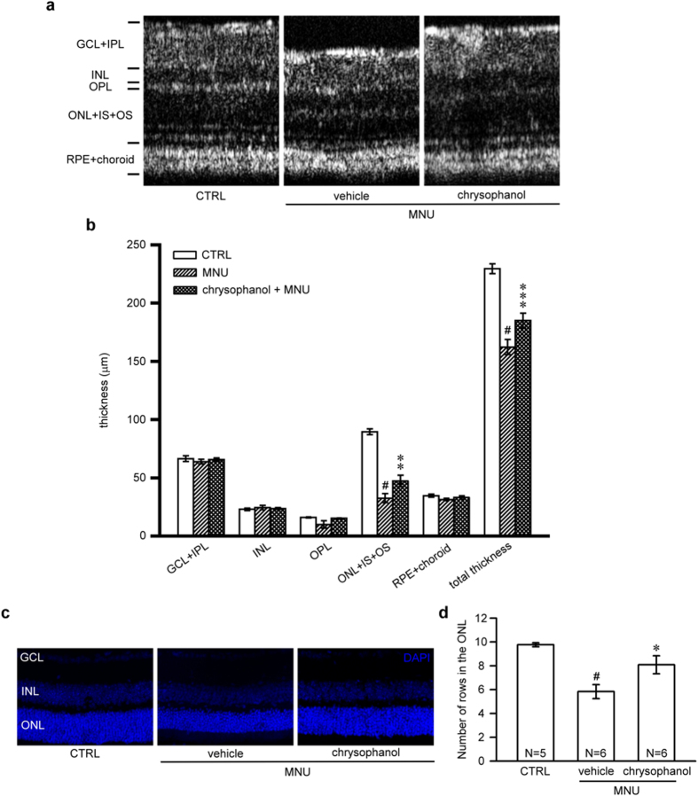
Figure 3. Chrysophanol attenuated MNU-induced photoreceptor degeneration on day 7.
(a) OCT was used to evaluate changes in retinal thickness in control mice (n = 7) and in mice treated with the vehicle control (n = 6) or chrysophanol (n = 8) 7 days after the administration of MNU (60 mg/kg). (b) Quantification of the thickness of the separated layer using InSight XL software. (c) The arrangement of nuclei in the GCL, INL and ONL in DAPI-counterstained retina sections. (d) Quantification of the number of photoreceptor nuclei rows in the ONL. The data are presented as the mean ± SEM. CTRL: control; N: number of animals in each group; GCL: ganglion cell layer; IPL: inner plexiform layer; INL: inner nuclear layer; OPL: outer plexiform layer; ONL: outer nuclear layer; IS: inner segment; OS: outer segment; RPE: retinal pigmented epithelium. #p < 0.001 compared with the control group treated with vehicle, *p < 0.05 compared with the MNU-exposed group treated with vehicle, **p < 0.01 compared with the MNU-exposed group treated with vehicle, ***p < 0.001 compared with the MNU-treated group treated with vehicle.

