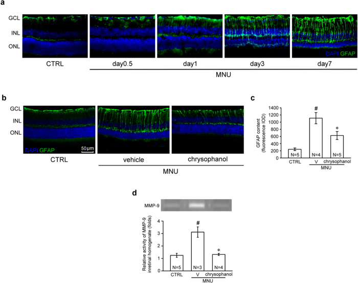
Figure 7. Chrysophanol inhibited MNU-induced reactive gliosis and retinal MMP-9 activation on day 7.
(a) The time course of GFAP localization (green) after MNU (60 mg/kg) administration. (b) GFAP content in DAPI-counterstained retinal sections. (c) MMP-9 gelatinolytic activity in retinal homogenates was detected using zymography. All the zymographic experiments were performed under the same conditions. After gelatin staining, we immediately cropped the gelatinolytic regions in the gels (Supplementary Fig. S2). The data are presented as the mean ± SEM. CTRL: control; V: vehicle; N: number of animals in each group; GCL: ganglion cell layer; INL: inner nuclear layer; ONL: outer nuclear layer; IOD: integrated optical density. #p < 0.001 compared with the control group treated with vehicle, *p < 0.001 compared with the MNU-exposed group treated with vehicle.

