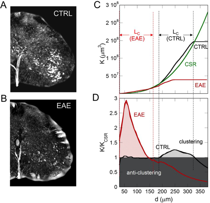
Figure 7.
Phase contrast tomographic volume, 700 μm thick of (A) healthy (CTRL) and (B) EAE samples. (C) K-Ripley function of the motor neurons calculated in a typical volume of (red line) EAE and (black line) CTRL samples. The thick green lines represent the K-Ripley function of randomly located neurons. (D) K/KCSR ratio, of the motor neurons calculated in the same volume of EAE (red line) and CTRL samples (black line). In the white region (ρ > 1) the neurons tend to clusterize within the range lengths, Lc, indicated by the dashed lines at 145 μm and at [185–330] μm in EAE and CTRL samples, respectively.

