
Fig. 5.
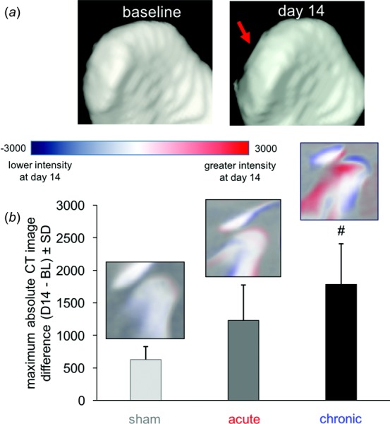
In vivo CT imaging of the TMJ showing changes in the overall joint architecture, with (a) more flattening of the TMJ condyle at day 14 with chronic pain than for sham controls and (b) quantification of changes in bone density. Changes in the CT image intensity at day 14 by image subtraction from baseline (BL) show the greatest change from controls in TMJ condyle structure with chronic pain (#p = 0.013).