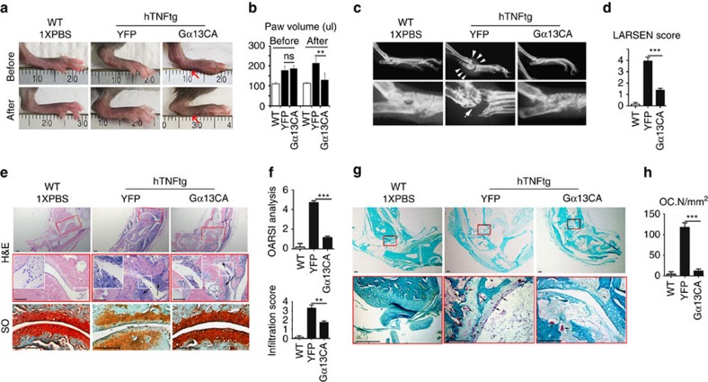
Figure 6. Gα13 protects Rhematoid arthritis mice from inflammatory bone loss.
(a–h) Histology analysis of 18-week-old male WT and hTNFtg Rheumatoid arthritis mouse ankles. hTNFtg mouse ankles were injected with AAV-Gα13CA or AAV-YFP. (a) Photographic images before and after AAV treatment. The red arrows showed paw swelling is relieved after AAV-Gα13CA treatment. (b) Quantification of hind paw volume in a; N=8. (c,d) Radiographic images; White arrows mark bone destruction. (d) Quantification of bone destruction (Larsen grade) in a; N=8. (e) H&E staining (black arrows mark monocyte infiltration) and Safranin O (SO) staining (black arrows mark articular cartilage damage). (f) Quantification of cartilage damage (OARSI grade) and inflammation (infiltration score) in e; N=8. (g) TRAP staining; black arrows mark TRAP positive cells. (h) Quantification of osteoclast number (Oc.N) in g; N=8. Results were expressed as mean±s.d.; *P≤0.05; **P≤0.01; ***P≤0.001 (Student's t-test). scale bars in e,g 200 μm.

