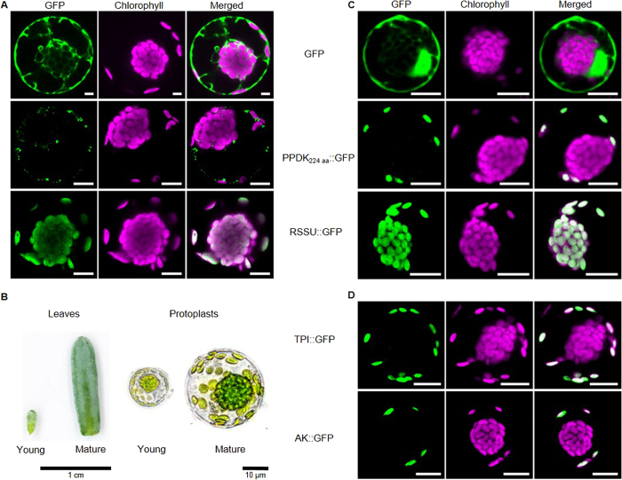
Figure 1. Subcellular localization of chloroplast targeted proteins in different developmental stages of Bienertia sinuspersici.
(A,C,D) Confocal images of various transiently expressed GFP-fusion proteins in B. sinuspersici chlorenchyma protoplasts. All fluorescence images are shown in the GFP channel (green - excitation 488 nm/emission 509/525 nm) and chlorophyll autofluorescence (red - excitation 408 nm/emission 620/700 nm). Additionally, the merged channels are shown. All images are representative from n ≥ 5 independent experiments. All scale bars = 10 μm. (A) Transient expression of GFP, PPDK224-GFP and full length RSSU-GFP in mature protoplasts. (B) Size comparison between young (Y) and mature (M) leaves and protoplasts. Scale bar leaves = 1 cm; Scale bar protoplasts = 10 μm. (C + D) Transient expression of GFP, PPDK224-GFP, RSSU-GFP, TPI-GFP and AK-GFP in young protoplasts. GFP – green fluorescent protein; PPDK – Pyruvate, Pi dikinase; RSSU – Rubisco small subunit; TPI – triosephosphate isomerase; AK – adenylate kinase.

