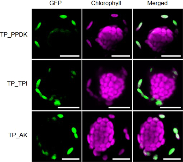
Figure 2. Subcellular localization of transit peptide GFP-fusions in young Bienertia sinuspersici protoplasts.

Representative confocal images for all tested GFP-fusion constructs (n > 5) of GFP-fusion proteins of the P-specific proteins PPDK, TPI and AK in chlorenchyma protoplasts (TP_PPDK, TP_TPI, TP_AK). Transit peptides length was predicted by ChloroP55 (Table S5). Protoplasts were analyzed as described in Fig. 1. Scale bars = 10 μm. GFP - green fluorescent protein; PPDK – Pyruvate, Pi dikinase; TPI – triosephosphate isomerase; AK – adenylate kinase.
