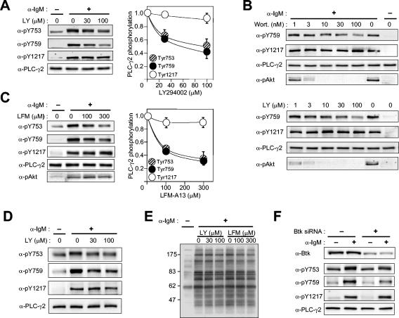
FIG. 6.
Effects of inhibition or depletion of Btk on BCR-induced PLC-γ2 phosphorylation in Ramos cells. (A) Effect of LY294002 (LY) on PLC-γ2 phosphorylation. Ramos cells were treated for 10 min with vehicle or the indicated concentrations of LY294002 before stimulation (or not) for 1 min with anti-IgM (25 μg/ml). Cell lysates were then subjected to immunoblot analysis with the indicated antibodies (α-) (left panels). The immunoblot intensities were corrected for the basal level of phosphorylation, normalized by the corrected value for stimulated cells pretreated with vehicle, and plotted against inhibitor concentration (right panels); data are means ± standard error of values from four independent experiments. (B) Effects of wortmannin (Wort.) and LY294002 on PLC-γ2 phosphorylation and Akt phosphorylation on Thr308. Ramos cells were treated for 10 min with the indicated concentrations of inhibitors before stimulation (or not) for 1 min with anti-IgM (25 μg/ml). Cell lysates were then subjected to immunoblot analysis with the indicated antibodies. (C) Effect of LFM-A13 on PLC-γ2 and Akt phosphorylation. Ramos cells were treated for 10 min with vehicle or the indicated concentrations of LFM-A13 before stimulation (or not) for 1 min with anti-IgM (25 μg/ml). Cell lysates were then subjected to immunoblot analysis with the indicated antibodies. (D) Effect of LY 294002 on PLC-γ2 phosphorylation in splenic B cells. Murine B cells were treated with indicated concentrations of the inhibitor for 10 min before stimulation for 1 min with anti-IgM (25 μg/ml). Cell lysates were then subjected to immunoblot analysis with the indicated antibodies. (E) Effects of LY294002 and LFM-A13 on overall tyrosine phosphorylation of cellular proteins. Lysates of Ramos cells similar to those in panels A and C were subjected to immunoblot analysis with antiphosphotyrosine. The positions of molecular size standards are shown in kilodaltons. (F) Effects of Btk depletion on PLC-γ2 phosphorylation. Ramos cells were transfected with a control RNA or Btk siRNA. Three days after transfection, the cells were left unstimulated or stimulated for 1 min with anti-IgM (25 μg/ml). Cell lysates were subjected to immunoblot analysis with the indicated antibodies.

