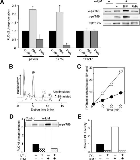
FIG. 7.
Effects of changes in PKC activity on the phosphorylation and activation of PLC-γ2 in Ramos cells. (A) Effects of BIM and PMA on PLC-γ2 phosphorylation. Ramos cells were incubated for 10 min in the absence or presence of 2.5 μM BIM or 1 μM PMA before stimulation (or not) for 1 min with anti-IgM (25 μg/ml). Cell lysates were then subjected to immunoblot analysis with the indicated antibodies (α-) (right panel). The relative blot intensities for stimulated cells (after correction for basal values) were determined as means ± standard error from three independent experiments (left panel). (B) Detection of [3H]inositol phosphates produced in unstimulated and BCR-stimulated cells. Ramos cells that had been metabolically labeled with myo-[3H]inositol were left unstimulated or were stimulated for 30 min with anti-IgM (25 μg/ml) in the presence of 20 mM LiCl. [3H]inositol phosphates in cell extracts were analyzed with an HPLC system equipped with an on-line radioactivity detector. (C) Time course of phosphoinositide hydrolysis. Cells labeled with myo-[3H]inositol were treated for 10 min with LiCl in the absence (closed circles) or presence (open circles) of 2.5 μM BIM and then stimulated for the indicated times with anti-IgM (25 μ/ml). [3H]inositol phosphates (IP + IP2 + IP3) in cell extracts were then measured as in panel B. (D and E) LY294002 sensitivity of the effect of BIM on BCR-induced PLC-γ2 phosphorylation (D) and on PLC activity (E). Ramos cells were treated for 10 min in the absence or presence of 2.5 μM BIM, 100 μM LY294002, or 2.5 μM BIM plus 100 μM LY294002 before stimulation for 1 min with anti-IgM (25 μ/ml). Cell lysates were subjected to immunoblot analysis with anti-pY759 (inset). The relative blot intensities were determined as means from three independent experiments. In panel E, cells labeled with myo-[3H]inositol were treated with BIM or LY294002 and then stimulated with anti-IgM as in panel D in the presence of 20 mM LiCl for 30 min. The amounts of [3H]inositol phosphates in cell extracts were then measured. Data are expressed as relative PLC activity after subtraction of basal values.

