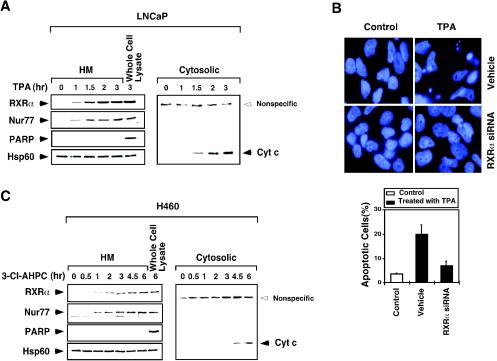
FIG. 2.
Time course analysis of mitochondrial targeting of RXRα and Nur77 and the release of cytochrome c from mitochondria. (A) Time course analysis of LNCaP cells in response to TPA. Cells were treated with TPA (100 ng/ml) for the indicated times. Mitochondrion-enriched HM and cytosolic fractions were prepared and analyzed for the presence of RXRα, Nur77, and cytochrome c as indicated. As a control, the whole-cell extract was also analyzed. Expression of mitochondrion-specific Hsp60 protein and nucleus-specific PARP protein was determined to control the purity of HM fractions. One of two similar experiments is shown. (B) Inhibition of RXRα expression suppresses the apoptotic effect of TPA. LNCaP cells were transfected with RXRα siRNA, followed by treatment with TPA (100 ng/ml) for 3 h. Cells were stained with DAPI and analyzed for nuclear morphological changes. Apoptotic cells were scored by examining 300 cells for apoptotic morphology from three different experiments. (C) Time course analysis of H460 cells in response to 3-Cl-AHPC. Cells were treated with 3-Cl-AHPC (10−6 M) and analyzed as described for panel A. One of two similar experiments is shown.

