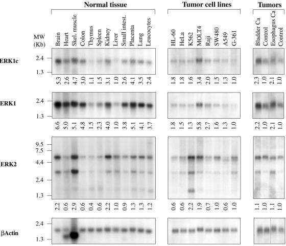
FIG. 2.
Distribution of ERK1c mRNA in cells and tissues. Northern blot analysis was performed on human tissues (MTN blots; Clontech), human cancer cell lines (MTN blots; Clontech), and human tumor blots (ResGen). The specific tissue, cell line, or tumor is indicated (Skel. muscle, skeletal muscle; Small intest., small intestine; Bladder Ca, bladder cancer). The probes used for detection were generated as described in Materials and Methods. The following probes were used: ERK1c unique sequence (ERK1c), human ERK1 N terminus (ERK1), human ERK2 N terminus (ERK2), and actin. Blots were hybridized in ULTRAhyb hybridization buffer (catalog no. 8670; Ambion) following the manufacturer's protocol. The positions of the RNA markers are indicated to the left of the gels. Relative expression was calculated by using a densitometer (model 690; Bio-Rad) and is indicated under the gels.

