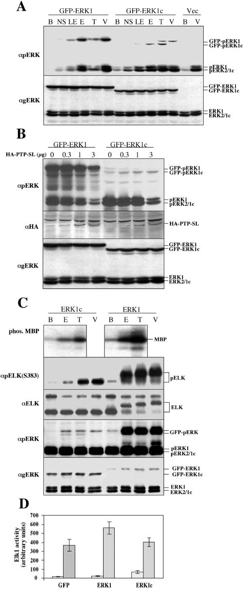
FIG. 6.
Biochemical properties of ERK1c. (A) Phosphorylation of exogenous ERK1c in response to extracellular stimuli. COS7 cells were transfected with GFP-ERK1 or GFP-ERK1c. Two days later, the cells were either not starved (NS) or the cells were starved and stimulated with either EGF for 14 h (10 ng/ml, 12 h) (LE), EGF (50 ng/ml, 10 min) (E), TPA (250 nM, 15 min) (T), VOOH (Na3VO4 [100 μM] and H2O2 [200 μM], 20 min) (V), or left untreated (B) as control. Cell extracts were analyzed by immunoblotting with the indicated Abs. Thedata are from a representative experiment (the experiment was performed three times). (B) Differential dephosphorylation of ERK1 and ERK1c by PTP-SL. COS7 cells were cotransfected with plasmids (1 μg each) containing either HA-ERK1 or HA-ERK1c, together with the indicated amounts of plasmid containing wild-type PTP-SL. After serum starvation, the cells were stimulated with EGF (50 ng/ml, 10 min) and harvested. Cytosolic extracts were subjected to an immunoblot analysis with the indicated Abs (anti-HA Ab [αHA] used to demonstrate increasing PTP-SL). The data are from a representative experiment (the experiment was three times). (C) Phosphorylation of MBP and Elk1 by ERK1c. COS7 cells were transfected with GFP-ERK1 or GFP-ERK1c. After serum starvation, the cells were stimulated with EGF (50 ng/ml, 10 min) (E), TPA (250 nM, 15 min) (T), or VOOH (18 min) (V) or left untreated (B). The GFP-ERKs were immunoprecipitated with anti-GFP Ab and subjected to an in vitro kinase assay with MBP or Elk1 as the substrate. The phosphorylation (phos.) of MBP was detected by autoradiography on an X-ray film (Agfa). The phosphorylation of Elk1 was detected by anti-pElk1(S383) Ab and also by upshift of Elk1 detected by anti-Elk1 Ab. The phosphorylation and amount of the GFP-ERKs were determined by an immunoblot analysis with the indicated Abs. The positions of phospho-MBP (pMBP), GFP-ERKs, GFP-phospho-ERKs (GFP-pERKs), and Abs are indicated at the sides of the gels. These results were from a representative experiment (the experiment was performed four times). (D) Effect of ERK1c on Elk1 transcriptional activity. HEK-293 cells were transfected with pFR-Luc (reporter), pFA2-Elk1 (fusion transactivator), pRenilla (reporter of transfection yield), together with GFP-ERK1 (ERK1), GFP-ERK1c (ERK1c), or GFP empty vector. Serum-starved cells were stimulated with EGF (50 ng/ml) for 14 h. Luciferase and Renilla luminescence were monitored as described in Materials and Methods. The results represent the means and standard errors (error bars) of three experiments.

