FIG. 8.
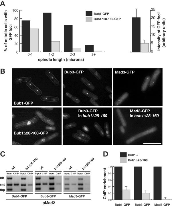
Bub1Δ28-160p fails to be efficiently enriched on central core chromatin and fails to recruit Bub3p and Mad3p to kinetochores. (A) Bub1p is recruited to kinetochores every cell cycle. bub1-GFP cdc25 cells were presynchronized in G2 at 36°C and then released at room temperature. Images of mitotic cells were captured after brief methanol fixation, and their spindle length was measured as the distance between two Cut12-CFP foci. Quantitation is displayed as the percentage of mitotic cells, with the defined spindle lengths, that displayed clear Bub1-GFP or Bub1Δ28-160-GFP foci. (B) Imaging of Bub1-GFP, Bub3-GFP, and Mad3-GFP in the indicated strains after their arrest in mitosis by Mad2p overexpression. While Bub1, Bub3, and Mad3 foci were readily detectable in the wild-type cells (70 to 80%) (upper panels), only faint Bub1Δ28-160-GFP foci could be imaged in living cells (∼20%) and Bub3-GFP and Mad3-GFP foci were undetectable in bub1Δ28-160 cells (lower panels). Bar, 10 μm. (C) ChIP demonstrated that there was reduced enrichment of central core DNA with Bub1Δ28-160-GFP and with Bub3-GFP, and almost no enrichment of Mad3-GFP, in bub1Δ28-160 strains that had been arrested by Mad2p overexpression. (D) ChIP quantitation. The DNA gels were scanned, and the enrichment of the ChIP signals, relative to the input, was calculated (50). We then set the enrichment for each protein in wild-type cells to a value of 100%. The reduced enrichment observed for each of the three proteins in the bub1Δ28-160 mutant was plotted as the percentage of this. This experiment was repeated four times, and the error bars show the maximum deviations from the plotted means.

